JuniperPublishers- Variation in Bifurcation of Brachial Artery: A Case Report
Journal of Surgery- JuniperPublishers
Abstract
Brachial artery, the main artery of the arm, usually distributes at the level of neck of radius into two branches. This article reports a case of high division of the brachial artery at the level of insertion of the coraco brachialis muscle. This variation, though not very rare, occurs in the embryo due to permanence of the upper portion of the radial artery rising from the brachial artery proximal to the origin of the ulnar artery followed by failure of development of the new connector of the radial artery with the brachial artery at the level of origin of the ulnar artery. High division of the brachial artery has a deep applied importance especially in the field of vascular surgery and radiology, and the feasibility of this variation should be bore in mind before any vascular surgery in the region of the forearm or while interpreting arterio grams of the upper limb.
Keywords: Brachial artery; Variation; Radial artery; Ulnar artery
Introduction
The brachial artery is the main artery of the arm and continuation of the axillary artery. Its proximal boundary is at the level of the inferior border of the teres major muscle and its distal boundary is at approximately a centimeter distal to the elbow joint at the level of the radial head. Passing below the bicipital aponeurosis, the brachial artery divides into the radial and ulnar arteries [1]. The artery is wholly superficial, covered anteriorly by skin and fascia and crossed superficially by the median nerve from the lateral to the medial side [2]. The brachial artery gives origin to profunda brachii, nutrient, superior and inferior ulnar collateral and muscular [3]. Variations in the arterial supply of the upper limb are relatively usual, with reported frequencies of incidence ranging from 11 to 24.4%. They can be found at different situations along the axillary, brachial, radial, or ulnar arteries, as well as in the palmar arches [4]. The recognition and documentation of expansion variation in the course, distribution and branching pattern of the arteries of upper limb is highly considerable in the angiographic and surgical practice [5]. In the present case, a rare variation of brachial artery apportions into radial and an ulnar artery in middle third of arm with superficial course of radial artery in right forearm is presented.
Case Report
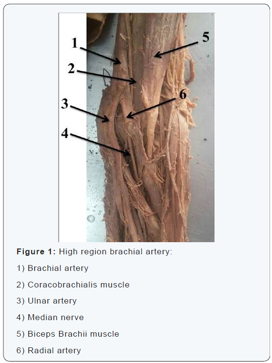
During anatomical dissection of the left upper limb of a male Iranian cadaver in the Medical Faculty of Tehran University of Medical Sciences, an anatomical variation of the high region brachial artery was recognized. The skin and fascia of the left upper limb was completely dissected, and the neurovascular bundles of the axillae and the whole limb was clearly explored. The course of the arteries and their branches were thoroughly traced and the observed anatomical variation was recorded. The axillary artery in left upper limb had normal anatomical course and branching pattern with the branches of brachial plexus cords normally distributed around it. The radial artery arose at the level of insertion of the coraco brachialis muscle. The brachial artery continued as ulnar artery in the forearm. Further course and branching of the radial and ulnar arteries in the forearm and palm was usual (Figure 1).

Discussion
In the upper limb sprout, the axis artery is derived from the lateral branch of the seventh intersegmental artery that is subclavian artery [6]. The proximal part of the main trunk of this artery forms axillary and brachial arteries and its distal part continues as anterior interosseous artery and deep palmer arch(6). Radial and ulnar arteries are last to become evident in the forearm from the axis artery, that is brachial artery. Initially, the radial artery arises more proximally than the ulnar artery. Then, it establishes a new connection with the main trunk at or near the level of the ulnar artery. The upper portion of its original stalk usually disappears to a large extent. To date, the reasons for the various prevalence rates of arterial variations in different geographical regions are not well understood.
Conclusion
Diagnostically, this variation may disturb the assessment of arteriography images and can have serious implications in orthopedic and vascular surgeries. Also, variations in the arterial of this region may be encountered during arteriography test, percutaneous brachial catheterization, and skin flap elevations from the arm or forearm [7-11]. Blood pressure, which is regularly measured in the arm in the brachial artery, is also influence when there are double arteries instead of a single brachial artery. On the other hand, accidental intraarterial injections into the brachial artery may lead to ample bleeding, thrombosis, and loss of the upper limb [8,12]. Finally, being superficial, the brachial artery can be mistaken for a vein and accidental injection of drugs in this artery may cause reflex vascular obstruction resulting in disastrous gangrene of hand.
To read more articles in Journal of
Surgery
For More Open Access Journals in Juniper Publishers


Comments
Post a Comment